Journal Name: Journal of Applied Microbiological Research
Article Type: Case Report
Received date: 11 December, 2020
Accepted date: 13 January, 2021
Published date: 20 January, 2021
Citation: Chacón SC, Candanedo PGC (2021) Canine Dirofilariasis in Panama First Cases Report Immunochromatography and Necropsy Diagnosis. J Appl Microb Res. Vol: 4 Issu: 1 (07-17).
Copyright: © 2021 Chacón SC et al. This is an open access article distributed under the terms of the Creative Commons Attribution License, which permits unrestricted use, distribution, and reproduction in any medium, provided the original author and source are credited.
Abstract
Background: Living beings have suffered with the climate changes that are affecting the planet in the latest decades. This has allowed the development of many vectors with the correspondent increase in their vector-borne diseases (VBD). D.immitis is a worldwide distribution parasite found in most American countries, but heretofore it was not recorded on the Panamanian isthmus.
Methods:We report three cases, one from Boca Chica, San Lorenzo district, and two from Puerto Armuelles, Barú district, all in Chiriqui province. These dogs arrived with serious cardiopulmonary symptoms and thrombocytopenia. A quick snap Quattro test was performed showing a strong positive result for D. immitis for all three. The Boca Chica and second Puerto Armuelles dogs were euthanized at the owners request and the first Puerto Armuelles patient died a few hours later. Hearts were fixed in formaldehyde and sent for an anatomic histopathology
Results:Thirty whole adult nematodes were found, 14 females and 16 males coming from Boca Chica; 43 adults, 18 females, 25 males as well as 11 immatures; and 59 adults, 22 femeales, 37 males as 16 immatures coming from the first and second Puerto Armuelles dogs, respectively. It is located in the pulmonary artery and right ventricle and atrium. From the Boca Chica canine an adult nematode was collected from the pericardium. The anatomy histopathology results show chronic myocarditis, chronic pericarditis, and enlargement of the right ventricle for Boca Chica’s patient, in addition, the pulmonary artery-vein endothelium, and inner space seems to be normal. The Puerto Armuelles anatomy histopathology studies are in progress.
Conclusion:We can conclude, starting with the observed cases, that D. immitis epidemiological research is necessary in Panama to ascertain the real prevalence, distribution, biology and pathogenesis of the nematode and its symbionte in this country.
Keywords
Dirofilaria immitis, Heartworm disease, Canines, Panama, Vector borne disease, Mosquitoes, Pathogenesis, First case report.
Abbreviations:VBD: Vector borne disease, AHS: American Heartworm Society, HVHP: Hospital Veterinario Healthy Pet, MDofChiriquí: Medical Diagnostics of Chiriquí, WBC: White Blood Cells.
Background
Living beings are chellenged daily by the climate changes that have affected the planet in the latest decades. Some Vector-Borne-Diseases (VBD) have emerged and re-emerged due to weather favoring the developing of their vectors [1]. Year after year higher temperatures are registered, which causes shorter biological cycles, increase of the generation quantities per year, and increase in the period when the vectors are present during the differents months of the year. The Republic of Panama has suffered the effects of the VBD for hundreds of years. Even before the canal construction by the United States of America, the French people suffered defeat when attempting construction of an interoceanic route due to the VBD, mainly those transmitted by mosquitoes in the Panamanian isthmus.
Canine Dirofilariasis or Heartworm disease, vector zoonosis whose etiologic agent in the american continent is Dirofilaria immitis (Spirurida: Onchocercidae), affects several tissues of domestic and wild dogs, cats, black bears, ferrets, lions, otters, ocelots, seals, and accidentally humans [2-5]. This is a worldwide disease with higher prevalence in tropical and subtropical areas [6], and it is being underestimated in Latin American countries, yet present in most of them with a few exceptions like Belice, Chile, Guatemala, Uruguay, French Guiana, and Panama [7]. This disease is confirmed in our neighboring countries Colombia [8] and Costa Rica [9-10] and has been described in littoraneals and continental areas, also happening in high altitude and cold weather locations [8]. The nematode is transmitted by mosquitoes of the genus Aedes, Culex, Anopheles, Culiseta and Taeniorhynchus [6,11]. The microfilariae or L1 circulating larvae are acquired by the mosquito after feeding on an infected definitive host. They develop between ten and fifteen days until transforming into L3 larvae, migrate to the insect’s mouth [6] and can infect a new definitive host in the next mosquito feeding. The L3 larvae are transmitted to the definitive host, in this case the dog, or to the accidental host, humans. The prepatent period varies from six to eight months. The L3, now in the dog’s blood stream, develops until L5 that migrate primarily to the pulmonary artery, secondarily to the right ventricle and pulmonary vessels, where they reach the sexual maturity and develop until adults between three to four months after reaching the circulatory system [12]. These parasites cause lesions on the vascular endothelium, congestion of the pulmonary artery and obstruction of the big vessels of the dog’s heart by adults filarias, alive or dead, making it difficult for blood to flow, finally leading to the right ventricle failure and liver congestion with hepatomegaly, dyspnoea, cough and fatigue [13]. Females are viviparus and release L1 larvae to the dog’s blood stream. In humans the parasite will not develop until the adult stage. Most of the L3 infectants larvae die at the bite location but some can migrate through the tissue, being the L4-stage, which forms subcutaneous granulomatous nodules between 60 and 120 days after penetration [6]. However, some L3 can reach blood capillaries, the right ventricle and branches of the pulmonary artery causing infarction in the lung parenchyma and forming coin-shaped granulomatous nodules often confused with neoplasms by the diagnostic imaging techniques [6,14-15]. This can occur as a benign disease, most of the time asymptomatic, causing cough, throat and chest pain, fever, fatigue, respiratory distress, shaking chills, myalgia, hemoptysis, and several bilateral pulmonary nodules with pleural effusion [6,16-18]. Misdiagnosis occurs due to the difficulty of recognizing the clinical form of the pulmonary Dirofilariasis and the potential of D. immitis to stay in other parts of the human body not just the lungs. Human exposure to D. immitis infective larvae is more often than registered in the different countries of Latin America [19].
New diseases emerge and others re-emerge in time, many transmitted by vectors, others by direct contact with infected animals. Aggravated by human intervention, the disease becomes each day a potential risk for animal health [20]. If we consider in addition to global warming, the increase in world population and the family migration with their pets, we can expect bigger chances of new cases of VBD appearing in the world. We have learned that it is of vital relevance to study the pathogens that affect the health of living beings. We live in an era where respiratory infections figure as major protagonists of deep scars in human history. It is crucial to determine the real impact of cardio-pulmonary parasites in the life quality of people and animals. We need detailed and current knowledge of all the elements that affect the delicate balance leading to an ideal state of health. The present article’s objective is to report three cases of Canine Dirofilariasis, one in Boca Chica, San Lorenzo district and two in Puerto Armuelles, Barú district, all in Chiriqui province, Republic of Panama.
Cases Description and Methods
The first case was observed in December 2019, was a male, bloodhound mixed breed weighing 28.5 kilograms. The dog was rescued when he was four months old from the streets of Boca Chica, which is located on the Pacific coast of the country in the San Lorenzo District, Chiriqui Province, Panama Republic (8º13’59.9’’N 82º13’59.9’’W). The dog had lived exclusively in Boca Chica until 5.5 years old. The pet was brought to our hospital, and the owner described the dog’s condition as showing total anorexia as well as a lack of liquid intake and convulsions for three days. The second case was observed in November 2020. An adult male, German Shepherd of 31.0 kg, bought at 3 months of age and introduced in the village of Puerto Armuelles, district of Baru, Pacific coast of the province of Chiriqui, Panama (8º16’39.9’’N 82º51’43.4’’W) where he lived until he was 3.5 years old. The animal arrived to the hospital with a history of not eating for seven days, agitation and aggression. The third case was observed in December 2020 coming also from Puerto Armuelles, was a two years old rescue female dog weighing 23 kg that came to us with anorexia, throwing up, lethargic and strong skin condition. In all cases an intravenous catheter was placed in the right cephalic vein to withdraw a blood sample and start fluid administration, and a hemogram and a quick immunochromatography tests were performed, Uranotest Quattro® Diagnostic Kit from Urano Vet for detection of Erlichia canis, Anaplasma platys and Leishmania infantum antibodies, and Dirofilaria Immitis antigens.
Results
During the physical examination of the Boca Chica dog we recorded a low temperature (37.8ºC); pale mucous membranes; congested, increased capillary fill time; prescapular lymph nodes bilaterally enlarged; weight loss; low body-score condition; increased in respiratory rate and abdominal breathing; an increase of heart impact; and severe arrhythmia. On physical examination, the first Puerto Armuelles patient presented increased prescapular lymph nodes, fever of 39.5ºC, congested mucous membranes, increased capillary perfusion time, on auscultation he showed arrhythmia, increased impact on the right side of the thorax, severe dyspnea with abdominal breathing. The second Puerto Armuelles patient showed enlarged prescapular and inguinal lymph nodes, corneal edema, serious cardiac arrhythmia and bradycardia, increased capillary perfusion time, normal temperature and severe dermatitis.
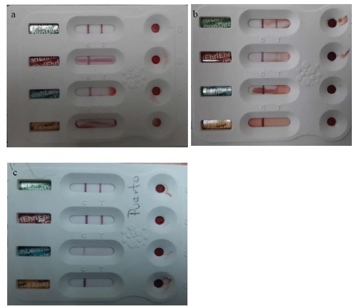
Uranotest Quattro® Diagnostic Kit showed positive result for D. Immitis antigens in all three cases but also positive to E. canis for both Puerto Armuelles sample (Figure 1 a,b,c). All hemogram results are detailed below (Table 1).
The Boca Chica dog’s hemogram shows an increase in the White Blood Cells (WBC) with absolute increase of monocytes and granulocytes, and a relative increase of eosinophils, microcytic hypochromic anemia, and thrombocytopenia. Due to the gravity of the convulsions, the low temperature and hemoglobin, the respiratory distress, and the Dirofilaria immitis positive diagnosis, the risk to human health, and the visible pet suffering, the owners requested for the dog to be euthanized. The procedure was performed using Xylazine 4.4 mg/Kg, Ketamine 20 mg/Kg and Potassium Chloride 100 mg/Kg [21].
Figure 1a, 1b, 1c:: Uranotest Quattro® Diagnostic Kit from Urano Vet. Canines’ whole blood positive to D. immitis from Boca Chica and Puerto Armuelles 1 and 2, respectively. Chiriquí, Panamá. Photo HVHP.
Both canines from Puerto Armuelles already presented relative lymphocytosis and thrombocytopenia. The second patient from Puerto Armuelles showed hypochromic anemia and a relative increase of eosinophils. The first patient from Puerto Armuelles showed symptoms of dehydration and weakness and died after a few hours later. The second Puerto Armuelles patient was euthanized by the owners’ request, due to the risk to human health, following the same procedure as for the patient from Boca Chica.
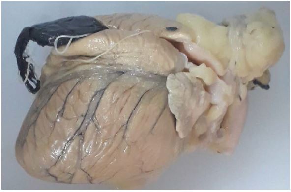
Hearts were extracted and fixed in formaldehyde for further histopathological anatomy observation (Figure 2).
Histopathological Anatomy Observations
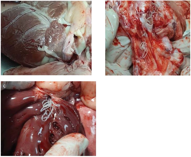
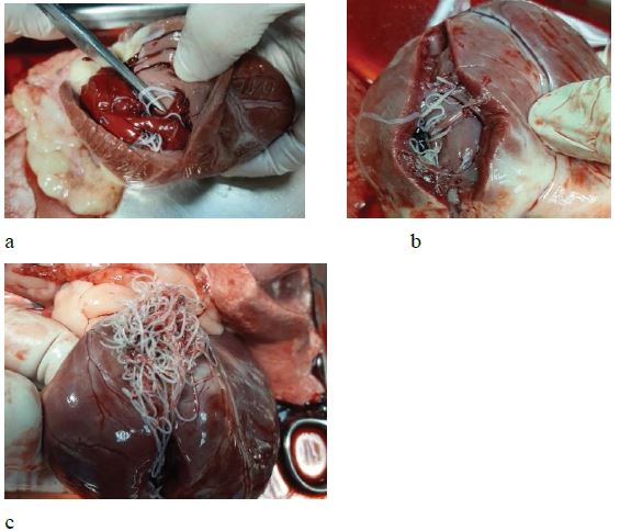
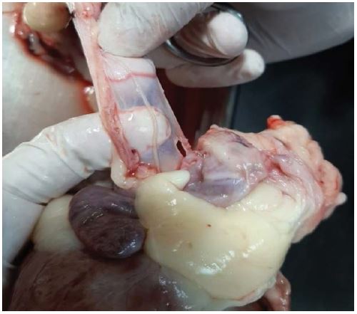
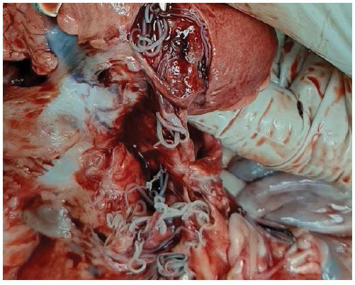
In all three patients D.immitis adults were observed in the pulmonary artery, in the right atrium and ventricle (Figures 3a,b,c and 4a,b,c); one nematode was observed in the pericardium of the Boca Chica dog (Figure 5). The pulmonary artery of the Puerto Armuelles first dog was full of nematodes which occluded a large portion of its lumen from the exit of the right ventricle until its arrival in the lung parenchyma (Figure 6). The nematodes in right ventricles of all the three patients were embedded in a large clot that filled all the ventricle light. The parasites were removed from the heart and clot.
Thirty adult whole nematodes were found, being 14 femeales measuring on average 20.2 cm (15-26.5) and 16 males measuring on average 12.4 cm (10-14.5) long (Figure 7) in the Boca Chica dog. No vegetation or atheromatous plaques were observed on the endocardium or great vessels.
Figure 2: Canine heart, from Boca Chica, Chiriquí, Panama, whole preserved in formaldehyde with adults of D. immitis emerging together with the clot through the cut in the right ventricle. Photo MDofChiriqui.
Figure 3a, 3b, 3c:Adults of D. immitis in the pulmonary artery lumen of canines from Boca Chica and Puerto Armuelles 1 and 2, respectively. Chiriquí, Panamá. Photo HVHP.
Figure 4a, 4b, 4c:Adults of D. Immitis in the right ventricle of canines from Boca Chica and Puerto Armuelles 1 and 2, respectively. Chiriquí, Panamá. Photo HVHP.
Figure 5:Adult of D. immitis in the pericardium of a canine from Boca Chica, Chiriquí, Panama. Photo HVHP.
Table 1: Whole blood Hemogram collected from canines positive for D. immitis and coming from Boca Chica and Puerto Armuelles. Chiriqui. Panama. Mindray BJC2800VET.
| Boca Chica case | Puerto Armuelles 1st case | Puerto Armuelles 2nd case | |
|---|---|---|---|
| WBC (6-17) | 26.3 x 10⁹/L | 8.0 x 10⁹/L | 8.6 x 10⁹/L |
| Lym# (1-5) | 5.0 x 10⁹/L | 3.5 x 10⁹/L | 2.9 x 10⁹/L |
| Mon# (0.15-1.35) | 1.9 x 10⁹/L | 0.4 x 10⁹/L | 0.7 x 10⁹/L |
| Gran# ((4-12.6) | 19.4 x 10⁹/L | 4.1 x 10⁹/L | 5.0 x 10⁹/L |
| Lym% (12-30) (2-9) | 19.1% | 43.2% | 33.6% |
| Mon% | 7.4% | 5.4% | 7.9% |
| Gran% | 73.5% | 51.4% | 58.5% |
| RBC (5.5-8.5) | 2.60 x 10¹²/L | 5.65 x 10¹²/L | 5.2 x 10¹²/L |
| HGB (12-18) | 2.7 g/dL | 13.3 g/dL | 11.0 g/dL |
| HCT (37-55) | 10.9% | 37.3% | 31.1% |
| MCV (60-75) | 42.1 fL | 66.1 fL | 59.9 fL |
| MCH (20-25) | 10.3 pg | 23.5 pg | 21.1 pg |
| MCHC (32-36) | 24.7 g/dL | 35.6 g/dL | 35.3 g/dL |
| RDW (11-15.5) | 20.7 % | 11.2 % | 14.6 % |
| PLT (200-700) | 115 x 109/L | 73 x 109/L | 162 x 109/L |
| MPV | 8.0 fL | 8.8 fL | 8.7 fL |
| PDW | 14.6 | 17.4 | 17.0 |
| PCT | 0.092% | 0.064% | 0.140% |
| Eos% (1-1.25) | 5.4% | 1.1% | 7.6% |
Figure 6:Adults of D. immitis occupying the lumen of the pulmonary artery and all the path to the lung parenchyma of a canine from Puerto Armuelles 1, Chiriqui, Panama. Foto HVHP.
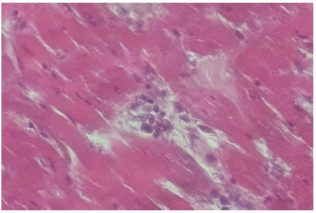
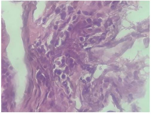
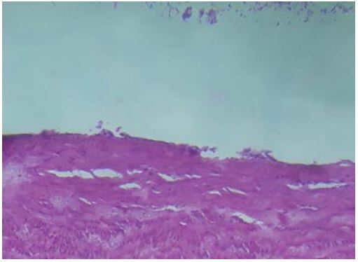
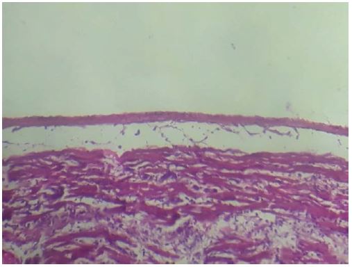
The coronaries were permeable. The pericardium had a transparent portion with one nematode adhered but without evidence of cardiac rupture, as if the parasite had developed there. No alterations were observed in the papillary muscles, tendinous cords or valves. Representative sections were made of the cardiac chambers, pulmonary artery and vein, and pericardium for microscopic assessment by histopathological evaluation. There was mild inflammatory infiltrate in the myocardium compound by lymphocytes and macrophages (Figure 8) and monocytes/macrophages infiltrate at the pericardium portion where the nematode was found (Figure 9). However, no macroscopic changes were observed such as opacity, thickening or stiffening. Endocarditis was not observed. The most relevant alterations of the heart were chronic myocarditis, mild right ventricle hypertrophy and chronic pericarditis. The evaluation of the light and the endothelium of the pulmonary artery were carried out using an endoscope, showing normality on both. The histopathological sections of the wall of the pulmonary artery and vein showed normality without alterations on the endothelium (Figures 10 and 11, respectively).
Figure 7:Adults of D. immitis removed by necropsy from the right ventricle and atrium, pulmonary artery and pericardium of a canine from Boca Chica, Chiriquí, Panamá. Photo HVHP.
Figure 8:Inflammatory Infiltrate composed of lymphocytes and macrophages in the myocardium of a canine from Boca Chica, Chiriquí, Panamá. Photo HVHP. Micrograph 200x, Hematoxylin-Eosin. Photo MDofChiriqui.
Figure 9:Monocyte and macrophage infiltrate in the portion of the pericardium where the nematode was found of a canine from Boca Chica, Chiriqui, Panama. Micrograph 200x, Hematoxylin-Eosin. Photo MDofChiriqui.
Figure 10:Histopathological section of the wall of the pulmonary artery of a canine from Boca Chica, Chiriqui, Panama, with normal appearance. Micrograph 200x, Hematoxylin-Eosin. Photo MDofChiriqui.
Figure 11:Histopathological section of the wall of the pulmonary vein of a canine from Boca Chica, Chiriqui, Panama, with normal appearance. Micrograph 200x, Hematoxylin-Eosin. Photo MDofChiriqui.
In the first patient from Puerto Armuelles, 43 adult nematodes were observed, with 18 females measuring 22.5cm (17-27cm) in length on average, 25 males measuring 13.5cm (11-16.0cm) and 11 immature nematodes measuring 8.3cm (5.5-11.5cm). In the second patient from Puerto Armuelles, 59 adult nematodes were found, with 22 females measuring 21.2cm (15.5-25cm) in length on average, 37 males measuring 13.9cm (10-16.5cm) and 16 immature nematodes measuring 12.1cm (8-15.5cm).The anatomy histopathological studies of Puerto Armuelles patients 1 and 2 are in progress.
Discussion
Heartworm disease caused by D. immitis, has been studied by various authors in the different countries of the world that make up the wide geographic distribution of the parasite. In Europe, they have studied the disease and concluded that the increase on its distribution is probably due to the increasing movement of infected dogs between countries, the increased interest to the disease, climatic changes, the emergence of new vector species and changes in the ecosystem due to human activities [22]. The study of high prevalence areas in Brazil registered that there is a tendency to find the highest percentages of positivity for D. immitis in areas where nature is better conserved [23]. In Colombia, the monitoring of some diseases transmitted by vectors to dogs in three cities of the country demonstrated a low prevalence of D. immitis in these regions, recommending constant monitoring and studies for the collection of samples and data to determine if the prevalence of this disease is increasing in this region [8]. In the United States, they developed a model to measure the risk of Dirofilariasis using some parameters such as temperature, precipitation, presence of forests, water surface coverage, presence of different species of mosquitoes, among others; therefore they concluded that all regions of the lower 48 states of the Union present some risk for infection by D. Immitis [24]. Also in the United States, a significant increase in the incidence of D. immitis in dogs was reported in the Southeast region, with an increase above the national average between 2013 and 2016 [25]. Canada reports a low incidence of the disease when compared to Borreliosis by Borrelia burgdorferi between 2013 and 2014 [26]. In Central America, more specifically in Nicaragua, its prevalence in dogs was found to be low-1.8%, when compared to E. canis, 62.9%. On the other hand, in Costa Rica, serum prevalence for D. immitis was detected in 4.42% of the canines and it is highlighted that these animals constitute reservoirs for the infection of humans [27,10]. Outside continental lands, the Canary Islands are considered as a hyperendemic area for canine heartworm disease. Four of the seven islands presented high serum prevalence among their inhabitants (6.2– 12.7%) for human pulmonary Dirofilariasis. These islands also correspond to the highest prevalences for canine Dirofilariasis and with ideal conditions for the development of the vectors [28]. Maggi and Krämer 2019 [7] conducted a complete review of the occurrence of VBD in pets in Latin America and reported the absence of D. immitis registration in the Republic of Panama.
The commercial characteristics of the Interoceanic Isthmus are to provide services, with ease for international transactions and transportation of merchandise between countries. That would also make possible the international transit of some vectors or host. In addition, the Panamanian interior is very touristy, highly visited by immigrant foreigners seeking calm lands to enjoy their retirement and ultimately move to the country, often with their pets. Descriptive epidemiological studies of travelers in the United States and Europe establish that Dirofilariasis is an emerging parasitic disease of dogs and humans, and that there is a greater risk for travelers suffering from it. They also report that global warming has extended the transmission cycles of mosquito vectors, the enzootic distribution, and the prevalence of canine microfilariasis to regions previously considered non endemic [12]. Evidence exists of Dirofilariasis cases imported from one country to another [29-30]. As Panama is a country with a high amount of transit of foreigners and their pets, it is incumbent to recommend that tests of diagnosis for heartworm on the sanitary surveillance protocols be performed or required from the country of origin.
It is important to highlight that the activity exerted by the host is fundamental to define the degree of exposure to the risk of contracting the parasite. Thus, dogs used for hunting have a higher risk of contracting the disease, a risk that increases with the time that the animal practices the activity [31].
The necropsy of four dogs that died with strong clinical signs of D. immitis showed lesions in all organs causing circulatory disorders due to severe parasitism and the toxic action of the parasite. Adult parasites inserted in a large thrombus in the lumen of the right ventricle were observed, also verifying its dilation. The authors also reported that the erosion of the vascular endothelium of the pulmonary artery and the proliferation of structures with the appearance of pseudovilli, are the result of the mechanical action of the parasites in the vascular lumen [32]. The evaluation of the damage caused exclusively by D. immitis to the different organs of the canine becomes difficult to carry out in vivo, since normally the domestic canine, and mainly the domestic stray or rescue dog, suffers from other vector pathologies transmitted mainly by ticks such as Ehrlichiosis, Anaplasmosis, Borreliosis and Babesiosis among others, which also cause damage to the various dog tissues.
Studies show that D. immitis maintains a symbiotic relationship with a bacterium of the genus Wolbachia sp which is known to be of vital importance for the nematode [33]. Some research questions the degree of participation of the bacteria in the pathogenesis of the disease [34,35], others reported that this relationship (nematode-bacteria) must be studied to understand the pathological mechanisms that both agents provoke in the host’s organism [36].
Some authors report that Dirofilariasis due to D. immitis is an emerging zoonosis that has been neglected and underregistered in Latin American countries. Others comment that the quantification of entomological parameters that affect the risk of transmission of a vector disease to a population of susceptible hosts is of fundamental importance [12,37-38]. On the other hand, the entity that draws up the guidelines for the Heart Worm in the United States (AHS), blames climatic changes and animal mobilization, for the increase in the potential of infection of the Dirofilariasis. They also report that the urban development of non-endemic areas and areas of low incidence have caused the expansion and increase in the prevalence of the disease, since, in one way or another, this behavior determines the predisposition to accumulate water, either by the supply for urban areas or by the incorporation of agricultural techniques such as irrigation for plantations, which causes the expansion of the habitat of its vectors [39]. Researchers recommend including D. immitis within the differential diagnoses of respiratory pathologies and pulmonary nodules in humans [6,40].
Tourism has increased in the last decades in Panama. Inland regions of the country are highly sought by foreigners due to the preservation of their natural conditions, flora, and fauna. This tourist demand and economic opportunity have led to the invasion of preserved areas by projects often disclosed outside the country. The Republic of Panama presents ideal conditions, such as diversity of mosquitoes, preserved vegetation areas, settled foreign immigrants, ecological tourism, the effect of climate changes, among others, which leads us to conclude that Dirofilaria immitis is a parasite that can be very present in this country, this is the reason why we recommend exhaustive studies and multidisciplinary efforts to elucidate the real impact of this nematode and its symbionte in the national territory, as well as prevention and monitoring policies that help to hinder and control the invasion of VBD to the Panamanian isthmus.
Recommendations and Comments
We recommend policies to monitor imported animals through tests that prove negative results when entering the country through any border as well as the appropriate prevention and education programs for the population and their pets.
Conclusion
We can conclude, from the observed cases, that epidemiological studies are necessary for Dirofilaria immitis in the Republic of Panama to trace the real prevalence and pathogenesis of the nematode in this country, the identification of its vectors, the damage caused to the canine, the prevalence in wild animals, and the incidence of lesions in humans, in addition to studies that identify the presence of Wolbachia sp in the nematode that will clarify its participation in the pathogenesis of the disease.
Declarations
Ethics approval and consent to participate
There are no ethical implications or limitations in the present study. We do not deal with experimental animals. The study reports the first finding of Heartworm in a natural infected dog. The euthanasia was performed as an act of humanity due to our ethical commitment as a Veterinarian against the suffering of the animal, and as stipulated by the Panamanian law 133, March 2020.
Consent for publication
Not applicable.
Availability of data and materials
The datasets supporting the conclusions of this article are included within the article.
Competing interests
The authors declare that they have no competing interests.
Funding
Not applicable
Author´s contribution
Both authors made substantial contributions to the manuscript. SCCh and PGCC performed the clinical examination, laboratory tests and necropsy. SCCh wrote the manuscript. PGCC made corrections to the text. Both authors read and approved the final manuscript.
Acknowledgement
To my patients as they offer me the casuistry and experience necessary for correct understanding and exercise of veterinary medicine.
To our friends from Medical Diagnostics of Chiriquí (MDofChiriquí), Pathology Laboratory, Dr. Rolando Alvarado Anchisi, Pathologist, Hospital Chiriquí, Chiriquí, Panamá, for the anatomy histopathological evaluation.
To our Friends Susan Kelley and Giannina Espinoza, English teachers, for the corrections in the text.
Beugnet F, Chalvet-Monfray K (2013) Impact of climate change in the epidemiology of vector-borne diseases in domestic carnivores. Comp Immunol Microb Infect Dis 36: 559-566. [ Ref ]
Alho AM, Marcelino I, Colella V, Flanagan C, Silva N, et al. (2017) Dirofilaria immitis in pinnipeds and a new host record. Parasit Vectors 10: 142. [ Ref ]
Pereira BB, Bastos BF, Keidel L, Leles D, Brener B (2018) Feline hearthworm (Dirofilaria immitis) infection: first case report of serological diagnosis in Brazil, confirmed by molecular assay. An Acad Bras Cienc 90(S1): 2293-2297. [ Ref ]
Montoya-Alonso JA, Carretón E, García-Guasch L, Expósito J, Armario B, et al. (2014) First epidemiological report of feline heartworm infection in the Barcelona metropolitan area (Spain). Parasit Vectors 7: 506. [ Ref ]
Panã D, Rãdulescu A, Mitrea IL, Ionita M (2020) First report on clinical feline heartworm (Dirofilaria immitis) infection in Romenia. Helminthologia 57: 49-56. [ Ref ]
Klinge SEM, Robayo CP, Barreto MAC (2011) Dirofilaria Immitis: una zoonosis presente en el mundo. Rev Med Vet 22: 57-68. [ Ref ]
Maggi GR, Krämer F (2019) A review on the occurrence of companion vector-borne diseases in pet animals in Latin America. Parasit Vectors 12: 1-37. [ Ref ]
McCown ME, Monterroso VH, Cardona W (2014) Surveillance for Ehrlichia canis, Anaplasma phagocytophilum, Borrelia burgdorferi and Dirofilaria immitis in dogs from three Cities in Colombia. J Spec Oper Med 1: 86-90. [ Ref ]
Rojas A, Rojas D, Montenegro VM, Baneth G (2015) Detection of Dirofilaria immitis and other arthropod-borne filaroids by an HRM realtime qPCR, blood-concentrating techniques and a serological assay in dogs from Costa Rica. Parasit Vectors 8: 170. [ Ref ]
Springer A, Montenegro VM, Schicht S, Vrohvec MG, Pantchev N, et al. (2019) Seroprevalence and Current Infections of Canine Vector-Borne Diseases in Costa Rica. Front Vet Sci 6:164. [ Ref ]
Diaz JH (2015) Increasing Risks of Human Dirofilariasis in Travelers. J Travel Med 22: 116-123. [ Ref ]
Silva CR da, Langoni H (2009) Dirofilariosis. Zoonose Emergente Negligenciada. Cienc Rural 39: 1614-1623. [ Ref ]
Acha PN, Szyfres B (2003) Filariasis zoonóticas. Zoonosis y enfermedades transmisibles comunes al hombre y a los animales. OPS, Washington. [ Ref ]
Simón F (1991) A seroepidemiologic survey of human dirofilariosis in western Spain. Trop Med Parasitol 42: 106-108. [ Ref ]
Simón F, Kramer LH, Román A, Blasini W, Morchón R, et al. (2007) Immunopathology of Dirofilaria immitis infection. Vet Res Commun 31: 161-171. [ Ref ]
Amato Neto V (1993) Dirofilariose pulmonar humana, adquirida no Brasil: comunicação de um caso. Rev Inst Med Trop Sao Paulo 35: 457-460. [ Ref ]
Cavallazzi RS (2002) Dirofilariose pulmonar humana: relato de sete casos. J Pneumol 28: 100-102. [ Ref ]
Vital RJ (2006) Human pulmonary dirofilariasis: atypical presentation of a rare disease. Rev Soc Bras Med Trop 39: 94-95. [ Ref ]
Theis JH (2005) Public health aspects of dirofilariasis in the United States. Vet Parasitol 133: 157-180. [ Ref ]
Santos Júnior EL dos, Moura JCR, Protásio BKF, Parente VAS, Veiga MHND (2020) Clinical repercussions of Glanders (Burkholderia mallei infection) in a Brazilian child: a case report. Rev Soc Bras Med Trop 53: e20200054. [ Ref ]
Leary SL, Underwood W, Anthony R, Cartner S, Corey D, et al. (2013) AVMA guidelines for the euthanasia of animals. [ Ref ]
Morchón R, Carretón E, González-Miguel J, Mellado-Hernández I (2012) Heartworm disease (Dirofilaria immitis) and their vectors in Europe – new distribution trends. Front Physiol Syst Biol 3: 196. [ Ref ]
Labarthe NV (2014) Updated canine infection rates for Dirofilaria immitis in áreas of Brazil previously identified as having a high incidence of heartworm-infected dogs. Parasit Vectors 7: 493. [ Ref ]
Bowman DD, Liu Y, McMahan CS, Nordone SK, Yabsley MJ, et al. (2016) Forecasting United States heartworm Dirofilaria immitis prevalence in dogs. Parasit Vectors 9: 540. [ Ref ]
Drake J, Wiseman S (2018) Increasing incidence of Dirofilaria immitis in dogs in USA with focus on the southeast región 2013 – 2016. Parasit Vectors 11: 39. [ Ref ]
Herrin BH, Peregrine AS, Goring J, Beall MJ, Little SE (2017) Canine infection with Borrelia burgdorferi, Dirofilaria immitis, Anaplasma spp. and Erlichia spp. in Canadá, 2103-2014. Parasit Vectores 10: 244. [ Ref ]
Springer A, Montenegro VM, Schicht S, Pantchev N, Strube C (2018) Seroprevalence and current infections of canine vector-borne disease in Nicaragua. Parasit Vectors 11: 585. [ Ref ]
Cabrera ED, Carretón E, Morchón R, Falcón-Cordón Y, Falcón-Cordón S, et al. (2018) The Canary Islands as a model of risk of pulmonary dirofilariasis in a hyperendemic área. Parasitol Res 117: 933-936. [ Ref ]
Fuehrer HP, Auer H, Leschnik M, Silbermayr K, Duscher G, et al. (2016) Dirofilaria in Humans, Dogs, and Vectors in Austria (1978-2014)-From Imported Pathogens to the Endemicity of Dirofilaria repens. PLoS Negl Trop Dis 10: e0004547. [ Ref ]
Sabũnas V, Radzijevskaja J, Sakalauskas P, Paulauskas A (2019) First report of heartworm (Dirofilaria immitis) infection in an imported dog in Lithuania. Helminthologia 56: 57-61. [ Ref ]
Orr B (2020) Pig-hunting dogs are an at-risk population for canine heartworm (Dirofilaria immitis) infection in Eastern Australia. Parasit Vectors 13: 69. [ Ref ]
Pasca SA, Acatrinei D, Oprean OZ, Lazar M (2012) Vascular, hepatic and renal lesions by Dirofilaria immitis invasión in dogs. Arq Bras Med Vet Zootec 64: 841-846. [ Ref ]
Rossi (2010) Effects of doxycycline on the endosymbiont Wolbachia in Dirofilaria immitis (Leydi, 1856) – Naturally infected dogs. Vet Parasitol 174: 119-123. [ Ref ]
Dingman (2010) Association of Wolbachia with heartworm disease in cats and dogs. Vet Parasitol 170: 50-60. [ Ref ]
Ceribasi AO, Simsek S (2012) Histophatology Effects of Dirofilaria immitis Microfilaria on Internal Organs of Dog Confirming by PCR Technique. Iranian J Parasitol 7: 103-107. [ Ref ]
Simón F, Siles-Lucas M, Morchón R, Gonzalez-Miguel J, Mellado I, et al. (2012) Human and Animal Dirofilariasis: the Emergence of a Zoonotic Mosaic. Clin Microbiol Rev 25: 507-544. [ Ref ]
Cirer AI, Rodríguez EB, Manzaba MJ, Gavilánez MC (2019) Clinical – Epidemiological update: human infection by Dirofilaria immitis and other zoonoticic filarias. J Scien Res 4: 1-17. [ Ref ]
Capelli (2013) Risk of canine and human exposure to Dirofilaria immitis infected mosquitos in endemic areas of Italy. Parasit Vectors 6: 60. [ Ref ]
American Heartworm Society (AHS) (2014) Current Canine Guidelines for the Prevention, Diagnosis, and Management of Heartworm (Dirofilaria immitis) Infection in Dogs. [ Ref ]
Montoya-Alonso JA, Morchón R, Cordón FY, Cordón FS, Simón F, et al. (2017) Prevalence of heartworm in dogs and cats of Madrid, Spain. Parasit Vectors 10: 354. [ Ref ]
