Journal Name: Journal of Clinical Case Reports and Trials
Article Type: Historical Review
Received date: 23 May, 2018
Accepted date: 02 August, 2018
Published date: 09 August, 2018
Citation: Musitelli S, Bertozzi MA (2018) A Brief Historical Survey of the Illustrations of the Positions of the Foetuses into the Womb. J Clin Case Rep Trials. Vol: 1, Issu: 1 (34-38).
Copyright: © 2018 Musitelli S, et al. This is an openaccess article distributed under the terms of the Creative Commons Attribution License, which permits unrestricted use, distribution, and reproduction in any medium, provided the original author and source are credited.
Historical Review
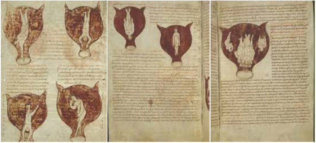
Neither any of the manuscripts of Hippocrates’ chapters dealing with gynaecology and paediatrics, nor Sorano’s of Ephesus’ (1st half of the 2nd century BC), Gynaikeia (Gynaecology) are provided with illustrations. A series of figures can only be found in Muscione’s (5th century AD), Gynaikeia, a Latin summary of Sorano’s Treatise preserved in the manuscript no 3714 (of the 9th century) of the Coninklijke Bibliotheek (or Bibliothèque Royale de Belgique) of Brusselles (Figure 1) [1].
Figure 1: The seven illustrations of the manuscript n. 3714 of the Coninklijke Bibliotheek (or Bibliothèque Royale de Belgique) in Brusselles.
However, nobody can avoid observing that:
- The womb is erroneously “horned” [1].
- The foetuses are not at all represented as they really are but like little and completely developed men. It is clear that the miniaturist had not even the faintest idea of a real foetus and did not absolutely draw from life!
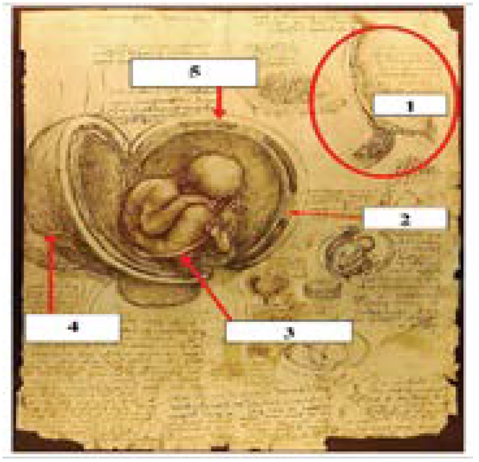
In order to find a realistic drawing of a foetus into the womb one must wait till Leonardo da Vinci’s (1452-1519) “Anatomical quaternions” (Figure 2).
Figure 2: Leonardo’s marvelous drawings of a foetus into the womb.
Figure 2 represents 1: the placenta; 2: the chorial villi; 3: the funiculus umbilicalis; 4: the uterine artery; 5: the uterine wall and its three layers. The drawings are surely a masterpiece, but it is worth observing that: A: the placenta is not a human, but a cow one; B: the position of the foetus is mistaken, unless Leonardo – who surely dissected a dead pregnant woman had the misfortune of dissecting a case of “breech presentation” (Partus agrippinus), which was generally deadly at his times [2]. However, the most important particular is that Leonardo represents the uterus as it really is instead of the “horned uterus” commonly described (From Hippocrates till the 15th century) and the consequent erroneous opinion, also advocated by Galen - that male offspring formed into the “right womb” and female offspring in the left one. These marvelous drawings were prepared by Leonardo as plates of a planned but never realized “Atlas of human Anatomy” he devised in Pavia “to provide [everyone] – as he writes in a marginal note - with a complete and correct anatomical knowledge”. This idea was surely conceived by Leonardo during his stay in Pavia as a consequence of his cooperation – most probably during the winter of 1510 – with the great Anatomist Marco Antonio della Torre (1481-15011), who was still holding – just in 1510, one year before his death, the chair of Anatomy at the University of Pavia.
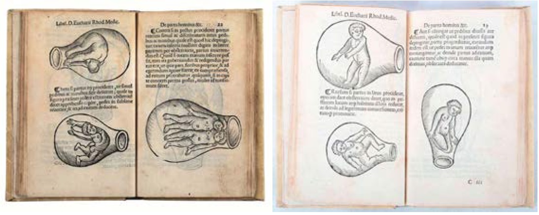
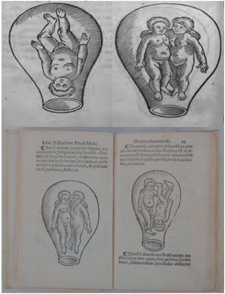
But let us now deal with the figures of Eucharius Rösslin’s (1470- 1526) “Rosengarten” (The garden of the roses) (Figure 3) and Scipione Mercuri’s (1540/50-c.1615) “La commare” (The midwife) (Figure 4) [3,4].
Figure 3: Rösslin’s figures: it is worth observing that although the womb is correctly not “horned”, nonetheless the foetuses are absolutely gimcrack drawings!
Figure 4: Four of Mercuri’s figures: also in this case it is worth observing that although the womb is correctly not “horned”, nonetheless the foetuses are absolutely gimcrack drawings!
Nothing different can be observed in Mercuri’s figures (Figure 4).
In Figure 4, the representations are nothing but completely developed babies a thick head of hair included! This means that neither Mercuri, nor Rösslin ever observed a real foetus into the womb!
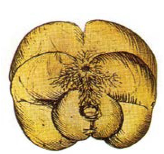
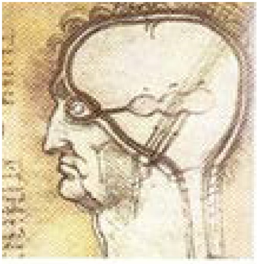
However, these evident absurdities are exceptionally interesting: they prove beyond any reasonable doubt that as Leonardo never published any of his anatomical drawings [5], he did not improve of even one only millimeter the anatomical knowledge. Moreover, in spite of some original observations, Leonardo confined himself to illustrating – as masterly as you won’t – what he read in the anatomical treatises of his time, big mistakes included. Suffice it to emphasize that he still draws the inexistent “rete mirabile” in the human brain (Figure 5) and not only did not distinguish the “dura mater” the “pia mater” and the “aracnoid”, but also represented the retina, the choroid, the sclera and the cornea connected with the brain meninges, a mistake already made by Galen and repeated by the subsequent anatomists till at last the 17th century (Figure 6).
Figure 5: The inexistent “rete mirabile” in the human brain but drawn by Leonardo on the basis of Mudinus’ erroneous description derived from Galen.
Figure 6: Leonardo’s illustration of a dissected human head. It is worth observing that the sclera and the cornea are erroneously connected with the meninges on the basis of Mundinus’ mistake derived, in its turn, from Galen’s erroneous statement.
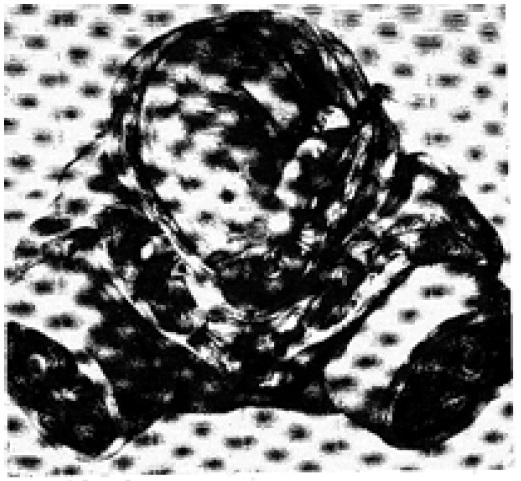
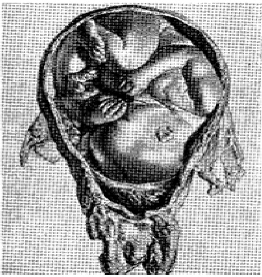
At last more than 1.200 years after Muscio and more than 1.500 years after Soranos of Ephesus one finds two perfect illustrations of a foetus into the womb in William Hunter’s (1718-1783) marvelous treatise “The anatomy of the gravid uterus”, printed by John Baskerville in Birmingham in 1774.
The treatise is provided with 34 perfect copper engravings.
However, we think that it is enough that one takes a glance of two of them (Figure 7 and Figure 8) to have an idea of the perfection of Hunter’s plates.
Figure 7: The first illustration of a foetus into the womb according to William Hunter.
Figure 8: The second illustration of a foetus into the womb according again to William.
Conclusion
There is no doubt that apart from Leonardo’s marvelous – although partially mistaken – drawings, nothing more perfect than Hunter’s plates could be realized even having recourse to the most sophisticated instruments we have nowadays at our disposal.
Acknowledgment
We could never interpret correctly Leonardo’s drawings (mistakes included) without the precious suggestions of our dearest friend Prof. Dr. Maurizio Claudio Bossi. We feel bound to express our special thanks to him.
KBR (1837) Koninklijke Bibliotheek van België–Bibliothèque royale de Belgique. Europeana Regia. [ Ref ]
Musitelli S and Bossi I (2016) Brief Historical Survey of Generation from Hippocrates (469-399 B.C.) to the Controversy between “Spermatists” and “Ooists”. Ann Reprod Med Treat 1: 1002. [ Ref ]
ER Rosengarten (1538) Latin translation, Johann Foucher, Paris. [ Ref ]
Mercuri S, La Commare o ricoglitrice, Venice GFB (1686). [ Ref ]
William Hunter (1774) The anatomy of the gravid uterus. John Baskerville, Birmingham. [ Ref ]
