Journal Name: Journal of Clinical Case Reports and Trials
Article Type: Research
Received date: 03 September, 2018
Accepted date: 11 September, 2018
Published date: 17 September, 2018
Citation: Kharbachi FZ, Haddad F, Tahiri M, Hliwa W, Bellabah A, et al. (2018) Abdominal Pelvic Mass Revealing a Giant Prostatic Adenocarcinoma. J Clin Case Rep Trials. Vol: 1, Issu: 2 (19-21).
Copyright: © 2018 Kharbachi FZ, et al. This is an openaccess article distributed under the terms of the Creative Commons Attribution License, which permits unrestricted use, distribution, and reproduction in any medium, provided the original author and source are credited.
Abstract
Prostate cancer is the most common cancer among men over 50 years old. It has a relatively good prognosis when diagnosed early, unfortunately in some cases the diagnosis is made at locally advanced stages. We report a case of a prostatic adenocarcinoma revealed by an abdominopelvic mass. 83-year-old patient with a right orchiectomy, admitted for a diffuse abdominal pain that has been developing for a month, associated to a constipation, urinary frequency and urgency. The abdominal examination found an extensive firm mass from the left iliac fossa to the hypogastrium. Ultrasound and abdominopelvic computed tomography (CT) showed a large, predominantly retroperitoneal abdominopelvic mass, additionally, showing no metastatic masses. The anatomopathological study of the trans-parietal biopsy of the mass as well as that of the prostate biopsy shows prostate glandular proliferation of prostatic PSA +. The dosage of PSA is 23,600 ng/ml. Consequently an Anti-androgen hormone therapy has been indicated.
Keywords
Abdominal-pelvic mass, Giant adenocarcinoma of the prostate, High PSA, Hormonothéraphie.
Introduction
Prostate cancer is the most common cancer in men over 50, it is exceptional before 40 years. It is the second leading cause of cancer death after lung cancer. It is a real public health issue.
Adenocarcinoma is the most common histological type, and its prognosis has improved considerably thanks to the various advances made in the diagnosis of this cancer in early stages in developed countries. However it’s insidious development has made unfortunately in several situations the diagnosis at locally advanced stages [1].
Giant adenocarcinoma of the prostate is a rare form of prostate cancer and causes differential diagnosis problems with different abdominopelvic masses [2].
Medical Observation
83-year-old patient admitted for an abdominopelvic mass. He had a right orchiectomy 8 years ago. The patient was admitted for diffused abdominal pain though the most intense in the left iliac fossa (LIF) and the hypogastrium without irradiation associated to an alternating diarrheaconstipation and urinary symptoms of pollakiuria, urinary burns and urinary urgency. The patient had a decline in general condition.
At his first admission, the patient was conscious, PS at 1. The abdominal examination found a firm extended mass from the LIF to the hypogastrium that has irregular contours and that is fixed to both superficial and deeper planes, measuring 17 × 13 cm, painless, without any inflammatory signs on the skin opposite nor murmurs identified during the auscultation. Perception during rectal examination of extrinsic compression at the anterior and right lateral wall of the rectum. No lymph nodes found.
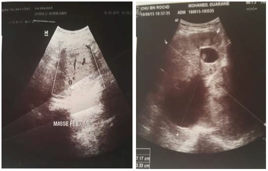
Patient underwent abdominal and pelvic ultrasonography showing a large heterogeneous echogenic solido-cystic mass that appears to have invaded the posterior wall of the bladder and extends to the left flank and left hypochondrium measuring 17 × 13 cm. It is responsible for a moderate left uretero-hydronephrosis. The prostate was not seen (Figure 1).
Figure 1: Ultrasound image of the abdominopelvic mass.
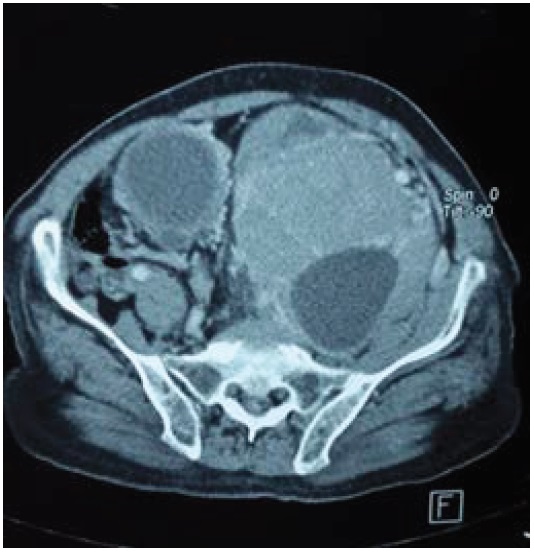
The abdominopelvic CT describes a large heterodense abdominal-pelvic mass with a multicomponent tissue, cystic regions and calcifications and irregular contours. It is predominantly retroperitoneal with detachment and repression in front of the primitive iliac vessels, measuring 15 × 13 × 19 cm.
This mass detaches the aorta though it remains permeable then comes in contact with the inferior vena cava which also remains permeable but with a partial overlapping and it engages the left iliac primitive vessels.
It settles in intimate contact with the bladder with which it has a blurry borders in some places. The bladder’s posterior wall is the site of an endoluminal thickening measuring 5 cm maximum (Figure 2).
Figure 2: CT image of the abdominopelvic mass.
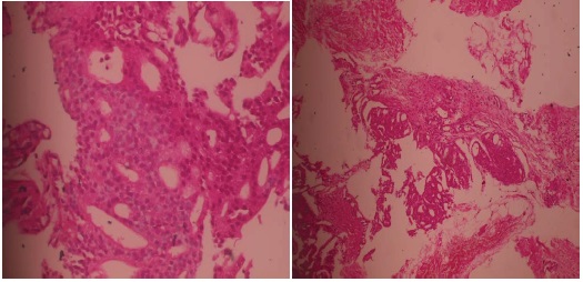
The exploration during cystoscopy found a multidiverticular muscular bladder. There was no evidence of mass or suspicious lesions in the bladder. Prostatic biopsy objectified an infiltrante prostate adenocarcinoma; score 5 according to GLEASON grade 3+2. The carrots examined are 40% invaded (Figure 3).
Figure 3: Histological aspect of the abdominopelvic mass in favor of prostatic adenocarcinoma.
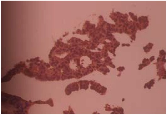
The transparietal biopsy of the mass also identified a glandular carcinomatous proliferation, with, at the immuohistochemical study, a positivity of PSA, a negativity of CK7, a negativity of CK20 (Figure 4). This aspect was in favor of a prostate glandular carcinoma proliferation.
Figure 4: Immunohistochemical study confirming the prostatic origin of the mass.
The PSA assay shows a rate of 23,600 ng / ml. An endoscopic digestive report (Oeso-gastroduodenal fibroscopy and colonoscopy) showed no abnormalities.
Discussion
In our patient’s case, the discovery of an abdominopelvic mass initially made us suspect a digestive cause. The urogenital origin was mentioned second due to his history of orchiectomy. A prostatic origin of the tumor was not thought of initially.
This clinical manifestation is not one of its kind. Kurokawa [2] has also reported a large abdominopelvic mass in a patient who also had a giant prostatic adenocarcinoma. It was the imaging and the histological findings that made the diagnosis of a prostatic origin of the abdominopelvic mass. Adenocarcinoma is not the only histological type of prostate cancer that can produce giant tumors. Indeed, other forms have also been described, in particular neuroendocrine prostate cancer, which can cause significant invasion of the gland [3].
A second case was also reported by Ze Ondo et al. a prostatic adenocarcinoma presented as a large hypogastric mass [4].
The clinical presentation of our patient was unconventional for a prostatic adenocarcinoma, neither the perception of an abdomino-pelvic mass extended from the left iliac fossa to the hypogastrium nor the digital rectal examination that found an extrinsic compression interesting the anterior and lateral wall or the transit disorder, thus the digestive origin of the mass was more probable, an endoscopic exploration was carried out, which proved to be normal.
The locally advanced stage of the tumor may explain the notion of urinary disorders reported by the patient. The biopsy of the abdominopelvic mass oriented the diagnosis towards a prostatic origin of the tumor, the rate of PSA was also very high. All these facts constituted a formal indication of prostate biopsy [5]. The thoraco-abdominopelvic computed tomography was sufficient for the extension assessment in our patient and did not objectify a secondary localization.
Therapeutically, anti-androgenic hormone therapy was indicated but not received by our patient who refused treatment and was confined to the family. Other methods of androgen suppression include surgical castration that can be performed by bilateral orchiectomy or sub-albuginea pulpectomy. The other methods are the agonists of the lutenizing hormone releasing hormone (LHRH), the antiandrogens and more recently the LHRH antagonists [1]. Hormone therapy can improve symptoms and reduce the risk of urinary and bone complications [6].
Conclusion
Abdominopelvic masses revealing prostatic adenocarcinoma remain rare. In our patient’s case, the radiology combined to the histological findings made the diagnosis possible. A thoraco-abdominopelvic CT did not show the existence of metastasis. Treatment with hormone therapy was indicated in this patient, who refused it.
There is no references


